
Doha Review: Al Ahli Hospital Ophthalmology Department
Brocade Blue
Monday, June 24, 2013
0 Comments


The ground floor houses among other departments; the emergency and pediatric units
I had a few vision tests done prior to the actual tests with Prof Alex. A female doctor, named Dr Dalia Khalid El-Faki ran the following tests:
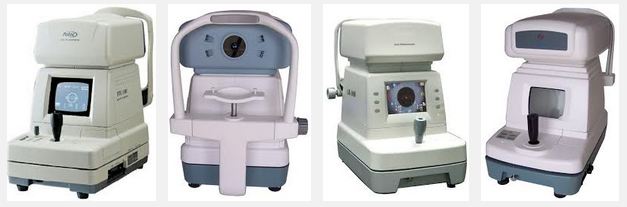
1. AUTOREFRACTOR/KERATOMETER test
Measuring my eyes' refractive error. An autorefractor was used to measures how light is changed as it enters my eyes, within seconds, it makes an approximate measurement of my prescription for eyeglasses or contact lenses and then prints it out
2. Visual Acuity Test
Measures the acuteness, clearness of my vision and the sharpness of the retinal focus within the eye and the sensitivity of the interpretative faculty of the brain

3. Cover Test
3.1 unilateral cover test
With my glasses on, I had to alternatively cover each eye with a handheld spoon-like device (scientifically known as the occluder) and focus on a dot (I forgot if it was red or blue) on the wall in front of me. Lights are turned off during this procedure. This procedure measures if I have strabismus or an eye turn/lazy eye
3.2 alternating cover test
I still had my glasses on, holding the occluder was switched from eye to eye, and had to read off letters from the wall across. This is done to observe if I have phoria. Although some amount of phoria is normal, large amounts can cause eye strain, blurry vision or double vision, as you must make an effort to fixate both eyes on a target

4. Digital phoropter
With that done, I was propped on a chair and with a digital phoropter working as my "glasses prescription machine" I was to read the last lines of letters on the eye chart in front of me, with each eye covered alternatively, and at the same time she changed the lenses and requested my feedback on which lense provided better vision. Finally, both the eyes were uncovered and I could focus much better on the chart. She inquired when I last had my eye test done, which was last year and she recommended me to get a new pair of glasses, because there was a slight increase in astigmatism in one of my eye. I was handed a new prescription for my eyes


Actual pictures taken from the hospital
After that was my appointment with Dr Alex. Like all Germans, their English is a mumble at best. He also looked like he truly enjoyed his job, whatnot with the jabbing and eye tests he ran Interestingly he was born in Indonesia but moved to Germany and after that to Middle East. He is the only ICO in the whole of Qatar, meaning if you go for your eye specialist exams he will be the judge. In Malaysia we two Indian doctors, one of each gender. Dr Alex also gets carried away with his test and forgets the patient is there unless you interact with him
So anyways the good doctor plainly refused to check my previous reading from Al Magrabi, stating that they have the two most expensive machines in their department which was the equivalent of Mercedes Benz in ophthalmology machines; named the Oculus Pentacam (conveniently made in Deutschland) Not only did it get my K1 and K2 readings, the curvature of my cornea, the thickness of my cornea but also my retina readings (front and back) This one machine did the work of probably 3 machines put together (pachymerty, topometry and retinoscopy) It also checks for early detection of keratoconus. If a patient shows a sign of this disease then LASIK is not recommended for him. Dr Alex does not perform LASIK himself, it is done by his wife, Dr Katharina, whom has had about 10 years in performing LASIK.
Prof. Dr. Alexander Arthur Bialasiewicz performed the following tests:
1. Tonometry
What is tonometry?
1.1. Goldmann Tonometry

The tonometry machine shown on their website, Dr Alex is on the right
This type of tonometry is the gold standard of taking precise and accurate intraocular pressure. It is most remembered for the blue light test. Yellow fluorescent dye was administrated into my eyes by an assistant. This thing burned my eyes. Apparently it helps patients to be able to see more, and it also has a numbing agent which helps the doctor to get a very very close look at your cornea, touching the tip of your cornea and flattening it to a certain point to check the pressure of the eyes (determined by the force applied to flatten it) The yellow dye kind of helps in reducing your eye blink count. I was instructed to focus at a blue dot in front of me. Of all the tests done, I'd have to say that I hate this machine the most because I had my eyes dilated twice for two types of tonometry! He also asked if I was born prematurely but he didn't explain why, but I told him I was born full term. Odd, to ask that. I don't know the medical reasons why but I'll ask my cousin if I see him around
2. Tomography with the Oculur Pentacam

Before he began the procedure the assistant administrated another dose of dilating liquid so I could get the best readings off the Pentacam
This was the one of the two most expensive device in their department, I am guessing the second one being the tonometry machine. The pentacam took all sorts of reading and did the work of three (or more probably) machines put together; pachymerty, topometry and retinoscopy.
After a few minutes of reading the paper, I was called back in for my final eye test
3. Applanation tonometry
By now my pupils were beginning to dilate nicely, albeit too slowly because again the assistant administrated another round of numbing drops and this one totally dilated my eyes. I was instructed to sit on the tonometry chair again for this procedure and roll my eye slightly up so he could place the probe. This is what Dr Alex said about the procedure; "it feels like deep sea diving only painful"

A special calibrated sterile probe is attached to a slit lamp biomicroscope is used to flatten part of the cornea. One end of the instrument is placed on the surface of the eyeball. This feels alot like having a heavy contact lens put in your eye, and after a few seconds I was struggling to keep my eye open and I could see the nerves at the back of my eye in my vision. Automatically, your reflexes tell you to move your head back and away from the chin rest and the forehead padded bar so the nurse held my head from the back to stop me from squirming too much.
After all my eye tests were completed, he sent me to his wife Dr Katharina whom sat down and explain the Pentacam eye reports to me (half of which I had forgotten but after spending a whole day on Google today I was able to read my own Pentacam reports)
Dr. Katharina Breidenbach
She started off by describing the disadvantages of LASIK, my eye conditions and recommendation for follow ups after the procedure. When I asked her if they do LASIK in AL Ahli, she said no they haven't got their machine yet so the false advertisement on their website has to go off. They have been waiting for their machine for over a year now. I wanted to know the differences with PRK and LASIK, so this is what she said:
1. In Comparison
PRK
A layer of cornea is removed, and this takes a longer time to grow back and is prone to infection. She has never done this type of surgery and doesn't recommend it for me because of my short-sightness and astigmatism. However, she has had patients whom has done this in the US and they come back to her with the common complain; that is they start seeing haze (blurry vision) after their surgery and they will probably be on life long need for eye drops
LASIK and any type of fancy-name-associated LASIK surgery
Involves removing the flap, lasering the tissue under the flap to get an accurate 20/20 vision the putting the flap back on. Because nothing is done to the flap, the tissue grows back to cover the gap, which happens overnight. Before I forget, she also mentioned a curable disease that attacks post-LASIK patients; known as the corneal ectasia
2. Reading the Pentacam Reports

2.1 The Scheimpflug image
The Scheimpflug image gives a complete representation of the anterior chamber, reaching from the endothelium to the posterior surface of the lens. Any opacities (tear, hole) of the cornea or lens are made visible and quantified objectively.

2.2 Topography maps of the anterior and posterior corneal surface
Previously it was impossible to get the readings from the back of the cornea, also known as the posterior eye readings and doctors only know the condition when they cut open the cornea flap. This machine does among other things:
- Keratoconus detection (you can't go for LASIK if you have this disease)
- Pre-surgical planning of refractive corneal surgery
- Follow-up after corneal surgery
- Calculation of IOL refractive power
- Planning of astigmatism reducing incisions (LRI)
- Follow-up after refractive surgery (pre-post LASIK)

2.3 Pachymetric
Remember the pen like pachymetry device that Al Magrabi used to check my cornea thickness? Here they used the Pentacam to do that
The corneal thickness progression analysis is calculated using concentric rings, starting at the thinnest point and extending to the periphery. Using this evaluation differences between normal corneas and Keratoconus corneas can be detected and used to identify very early changes. The results are displayed in two diagrams. For an objective comparison and analysis the progression index is calculated.
I'll explain what the graphs mean on the description in 2.8

2.4 Pachymetry maps, absolute and relative
It provides automatic representation of:
- corneal thickness in the centre of the pupil
- corneal thickness in the apex
- the thinnest point of the cornea
- corneal volume
In my case, my corneas are thick enough to undergo LASIK

2.5 Elevation maps of anterior and posterior corneal surface
This colour map serves to depict the elevation data of the corneal anterior and posterior surfaces using different reference bodies. It offers reliable assistance in the diagnosis of keratoconus, for ex., especially as elevation data are less prone to errors resulting from poor fixation by the patient than are curvature data.

2.6 3D anterior chamber analysis
This reading gives the automatic calculation of
- chamber angle
- chamber volume
- chamber depth

2.7 General overview display
This is the front page printed handout you get from the doctor
 The overview display provides important clinical information on the keratometry and pachymetry of the cornea in concise numerical form. The Scheimpflug image provides physician and patient with intuitive representations of opacities of the cornea or lens (cataract) or of the position of an IOL. The anterior chamber is described in terms of anterior chamber depth, volume and angle. When combined with IP tonometry readings corrected for corneal thickness the overall display permits an assessment of glaucoma risk. It also allows the display of all colour maps.
2.8 Overview display for refractive/LASIK surgeons
This is the overview page the doctor will be reading or going thru before hacking your eyes

As you can see from the left most curvature, my refraction is very close to the cross axis, 90-270 and 0-180 axis. This means they're not haywire, hence good for LASIK (if it was it wouldn't resemble a "t" but rather a motley of lines distributed all around the eye.
The top most graph shows the desired thickness of cornea and my cornea thickness is in red line, so its within the desired lines of 550-610mm. If it was an lesser than the top-most dashed line in blue; which is at 460 or 470 then the LASIK would probably give complications later after the surgery. When I asked Dr Katharina where would my thickness be after the surgery she said it would be somewhere along the dashed lines of 460/470-520/530mm
2.9 Power Distribution
The Corneal Power Distribution Display is a sophisticated tool to assess in detail corneal power. The Sim K’s, True Net Power and Total Corneal Refractive Power are displayed in a table chart

Based my blue spikes in the chart, you can see that it is only concentrated in a small region somewhere in the middle, and not distributed all over the graph. These are the K1 and K2 readings that you need before and after surgery because when you grow old and develop cataracts it is of extreme importance for a surgeon to calculate lens power using certain formulas. Otherwise it becomes a guessing mathematical game for him. Anyways this record is kept for eternity by your LASIK surgeon, so get him to give you a copy of the report. I have a detailed explanation on 2.10
2.10 True Net Power

Interestingly, Dr Katharina told me that the latest procedure for LASIK is the femtocesond LASIK and the machine is yet to come to Doha but when I checked Al Magrabi's website/my notes from Dr Ahmed he did mention acquiring the new machine for LASIK, it apparently has higher accuracy and recovery time.
The conclusion she gave based on my vision tests were that "I do not recommend you not to do LASIK" That's the closest to YES I got from her. Her responses were at best neutral when I requested her medical opinion if LASIK is do-able in Doha, or the guide to the right place to do it. But she did say that the latest machine is not yet available in Doha when Al Magrabi's Dr Ahmed clearly stated they have recently acquired it. I think that was her way of saying no, because she did mention that the weather here is dry, and LASIK will worsen it before it gets better. I went to Al Ahli with the intention of getting LASIK done there, and came out with a bunch of report from a fancy vision test machine.
My pupils were dilated and would stay that way for the next 4 hours, and I was having hard time adjusting my vision once I stepped out of the hospital, it felt like someone had exponentially increased the brightness and contrast of your laptop monitor. My vision was blurred & hazed so I dialed my husband and related the experience to him. I had to walk head down, with my umbrella covering my head and had to literally close my eyes to a Chinese level squint to hail a Karwa. Luck must be on my side because I got one in 5 mins. Then again maybe I wasn't so lucky, he didn't know the roads so I have to guide him with my GPS
Despite my momentary blindness I managed to get some pictures of the view from outside the hospital


The ophthalmology reception counter on 3rd floor


In conclusion, I am better off doing LASIK in Malaysia while I still have insurance coverage from Doha
References:
https://en.wikipedia.org/wiki/Ocular_tonometry
http://www.pentacam.com/sites/basic_features.php
http://www.pentacam.com/sites/hr_features.php
http://www.ebme.co.uk/arts/tonometry/
http://www.schuminweb.com/2012/10/11/two-days-two-eye-exams/


